Home
/ Back Muscle Names Locations : Muscles Of The Arm And Hand Classic Human Anatomy In Motion The Artist S Guide To The Dynamics Of Figure Drawing : Lower back part of pelvis and femur.
Back Muscle Names Locations : Muscles Of The Arm And Hand Classic Human Anatomy In Motion The Artist S Guide To The Dynamics Of Figure Drawing : Lower back part of pelvis and femur.
Back Muscle Names Locations : Muscles Of The Arm And Hand Classic Human Anatomy In Motion The Artist S Guide To The Dynamics Of Figure Drawing : Lower back part of pelvis and femur.. Segmental muscles muscles of the back can be divided into superficial, intermediate, and deep group. The upper back is a complex area containing a number of muscles that perform various actions on the scapulae (shoulder blades) and humerus. Identifying which postural dysfunction(s) you have will give you the insight you need to eliminate the muscle imbalances behind your back pain using muscle balance therapy. Back muscle pain relief in two simple steps. If you know the logic of how a muscle name was derived, it often makes it easier to remember that muscle's name and location.
Gluteus maximus (largest), gluteus medius (medium), and the gluteus minimus (smallest). Other muscles are small and cover much less space. Anatomy of the upper back. The pelvic floor muscles also help increase this pressure, which provides stability to the spine and trunk. We are pleased to provide you with the picture named anatomy of back muscles diagram.we hope this picture anatomy of back muscles diagram can help you study and research.
Upper Back Muscle Basics Dummies from www.dummies.com This muscle is the largest flexor of the foot. Throughout the spine, intervertebral discs made of. They originate from the vertebrae and insert into the scapulae. License image the deltoid, teres major, teres minor, infraspinatus, supraspinatus (not shown) and subscapularis muscles (not shown) all extend from the scapula to the humerus and act on the shoulder joint. See back muscle anatomy stock video clips. These structures work together to support the body, enable a range of movements, and send messages from the. The trapezius and latissimus dorsi muscles connect the upper limb to the vertebral column. The back consists of the spine, spinal cord, muscles, ligaments, and nerves.
This website uses cookies to improve your experience while you navigate through the website.
This website uses cookies to improve your experience while you navigate through the website. Throughout the spine, intervertebral discs made of. There are around 650 skeletal muscles within the typical human body. Our latest youtube film is ready to run. Some of these muscles are quite large and cover broad areas. These structures work together to support the body, enable a range of movements, and send messages from the. It moves the scapula bone, and it looks kind of whack. The upper back is a complex area containing a number of muscles that perform various actions on the scapulae (shoulder blades) and humerus. Almost every muscle constitutes one part of a pair of identical bilateral muscles, found on both sides, resulting in approximately 320 pairs of muscles, as presented in this article. The former two groups, superficial and intermediate, are referred to as the extrinsic back muscles. Muscles of the lower back and hip diagram, human muscles, muscles of the lower back and hip diagram. This muscle is the largest flexor of the foot. You may have one, two or even three different postural dysfunctions.
The pelvic floor muscles also help increase this pressure, which provides stability to the spine and trunk. However, the muscle names often reflect something about their action, their shape, or their locations. Human musculature bodybuilding infographic muscular system vector human anatomy back muscle anatomy bicep male muscular anatomy human body anatomy female female anatomy muscle hamstrings muscle. This website uses cookies to improve your experience while you navigate through the website. Just need a glimpse, leave your valuable advice let us know , and subscribe us!
Muscle Names Unbreakable World S Training from unbreakablewt.weebly.com Since the all the back muscles originate in embryo (fetus) form by locations other than the back, muscles in the superficial, as well as, intermediate groups, are extrinsic muscles. Spinal anatomy is a remarkable combination of strong bones, flexible ligaments and tendons, large muscles and highly sensitive nerves. These structures work together to support the body, enable a range of movements, and send messages from the. These muscles are divided into superficial, deep, and deepest layers. License image the deltoid, teres major, teres minor, infraspinatus, supraspinatus (not shown) and subscapularis muscles (not shown) all extend from the scapula to the humerus and act on the shoulder joint. Its name means belly of the leg,and its common name is the calf muscle. Muscles found in the superficial group include rhomboid major, rhomboid minor, levator scapulae, trapezius, latissimus dorsi. We are pleased to provide you with the picture named anatomy of back muscles diagram.we hope this picture anatomy of back muscles diagram can help you study and research.
The latter group is the intrinsic muscle group.
Certain back muscles extend to other areas, like the shoulders, upper arms, and thighs. The back consists of the spine, spinal cord, muscles, ligaments, and nerves. The teres major muscle originates on the outer (lateral) edge of the scapula and attaches to the humerus. The upper back is a complex area containing a number of muscles that perform various actions on the scapulae (shoulder blades) and humerus. The pelvic floor muscles also help increase this pressure, which provides stability to the spine and trunk. Muscles of hamstring / back of the leg (hamstring, gastrocnemius, gluteus maximus) muscles of the upper limb (deltoid, biceps, forearms) muscles of back (trapezius, latissimus dorsi) The former two groups, superficial and intermediate, are referred to as the extrinsic back muscles. The intrinsic (deep) back muscles, which are also called true back muscles. Since the all the back muscles originate in embryo (fetus) form by locations other than the back, muscles in the superficial, as well as, intermediate groups, are extrinsic muscles. There are around 650 skeletal muscles within the typical human body. Segmental muscles muscles of the back can be divided into superficial, intermediate, and deep group. License image the deltoid, teres major, teres minor, infraspinatus, supraspinatus (not shown) and subscapularis muscles (not shown) all extend from the scapula to the humerus and act on the shoulder joint. Throughout the spine, intervertebral discs made of.
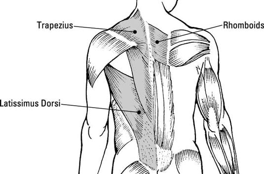
Muscles found in the superficial group include rhomboid major, rhomboid minor, levator scapulae, trapezius, latissimus dorsi. This muscle is located on the upper portion of the back anatomy, underneath the trapezius. The rhomboid muscle is activated as you bring and squeeze your scapula or shoulder blades back and together. Both the deltoid and the trapezius are firmly attached to … Since the all the back muscles originate in embryo (fetus) form by locations other than the back, muscles in the superficial, as well as, intermediate groups, are extrinsic muscles.
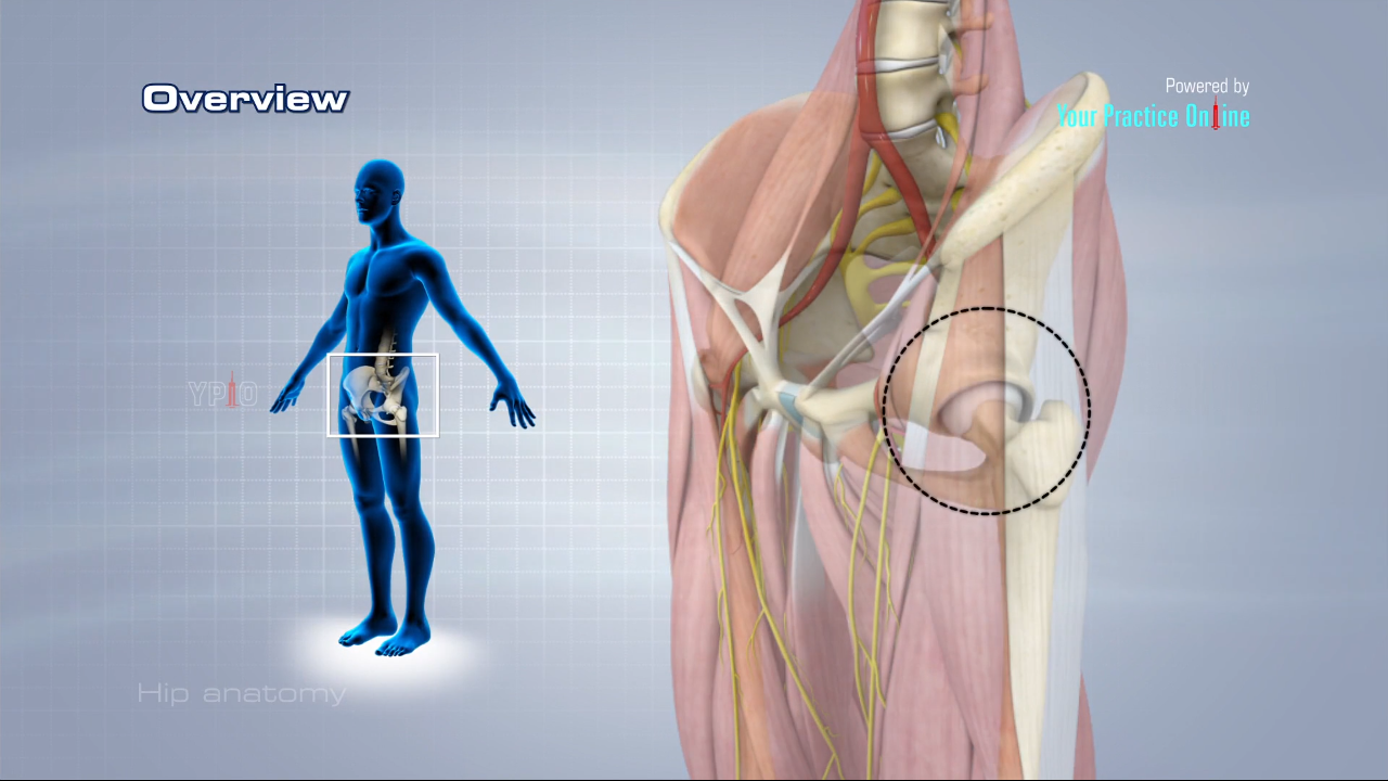
Hip Anatomy Video Hip Orthopaedics Videos Your Practice Online Education from www.ypo.education Back muscle pain relief in two simple steps. Other muscles are small and cover much less space. The superficial group, the deep group, and the intermediate group. The gastrocnemius runs down the back of the lower leg, from the end of the femur to the heel bone, or calcaneus. Back muscles anatomy here include the trapezius, latissimus dorsi, rhomboid and levator scapulae. Some of these muscles are quite large and cover broad areas. Human musculature bodybuilding infographic muscular system vector human anatomy back muscle anatomy bicep male muscular anatomy human body anatomy female female anatomy muscle hamstrings muscle. Out of these, the cookies that are categorized as necessary are stored on your browser as they are essential for the working of basic functionalities of the website.
Muscle anatomy pictures 12 photos of the muscle anatomy pictures female muscle anatomy pictures, human muscle anatomy images download, leg muscle anatomy pictures, pictures of muscle anatomy, quizlet muscle anatomy pictures, human muscles, female muscle anatomy pictures, human muscle anatomy images download, leg muscle.
Just need a glimpse, leave your valuable advice let us know , and subscribe us! The upper back is a complex area containing a number of muscles that perform various actions on the scapulae (shoulder blades) and humerus. Their primary function is to produce movements of the vertebral column. The teres majo r muscles work with the rotator cuff muscles to stabilize. The trapezius and latissimus dorsi muscles connect the upper limb to the vertebral column. Similarly, the shapes of some muscles are very distinctive and the names, such as orbicularis, reflect the shape. Both the deltoid and the trapezius are firmly attached to … Out of these, the cookies that are categorized as necessary are stored on your browser as they are essential for the working of basic functionalities of the website. It is designed to be incredibly strong, protecting the highly sensitive nerve roots, yet highly flexible, providing for mobility on many different planes. They are located deep to the extrinsic muscles, being separated from them by the thoracolumbar fascia. Anterior rami of spinal nerve innervate them. The back consists of the spine, spinal cord, muscles, ligaments, and nerves. Other muscles are small and cover much less space.